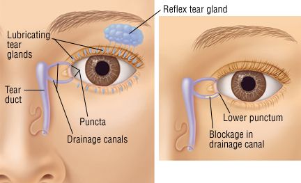
Tear Ducts Anatomy
The nasolacrimal duct is about 12 to 24 cm long. Tears consist of a complex and usually clear fluid that is diffused between the eye and the eyelid.
 Lacrimal Apparatus Includes Lacrimal Sac Gland Punctum Canaliculus Nasolacrimal Duct Inferior Meatus Of Nasal Cavity Jpg 580 Eye Skin Care Optometry Eye Facts
Anatomy and physiology of efferent tear ducts The human nasolacrimal ducts consist of the upper and lower lacrimal canaliculi the lacrimal sac and the nasolacrimal duct and drain tear fluid from the ocular surface into the nose.

Tear ducts anatomy. The second layer of the tear film is the aqueous layer and is secreted by the lacrimal glands which are located just below the eyebrow on the temporal near the temple side of the eyelid. The lacrimal gland produces tears. A baby can be born with a blocked tear duct a congenital blocked tear duct.
The diagnosis for Tear Duct Infection begins with physical examination of the eye. These are the small palpebral portion that lies closer to the eye and the orbital portion that forms around four ducts. Tears consist of a complex and usually clear fluid that is diffused between the eye and the eyelid.
The lacrimal gland lacrimal sac and nasolacrimal duct. A blocked tear duct also known as dacryocystitis happens when there is an obstruction in the passageway that connects the eyes to the nose or when the duct fails to open. A leading cause of blocked tear ducts in adults is infection of the eyes tear duct system or nasal passages.
Lacrimal gland is the gland that produces tears and the tears drain out onto the eye through two openings called the lacrimal ducts. This gland is about the size of an almond and sits within the lacrimal fossa located in the superior and outer edge of the orbital roofThe gland is divided into two sections anatomically. The tears then travel through the small canals in the lids canaliculi to a sac where the lids are attached to the side of the nose lacrimal sac then down a duct the nasolacrimal duct before emptying into your nose where they evaporate or are reabsorbed.
This duct runs through the eyes and into the nasal cavity it actually drains some of the excess tears into the inferior nasal meatus. Further components of the tear film include an inner mucous layer produced by specialized. Tear glands and tear ducts.
The nasolacrimal duct also called tear duct latin. Tear duct and glands. When the tear ducts are functioning correctly tears roll down from the lacrimal glands which sit above the outer side of each eye and across the surface of the eye.
It drains tears through the nasal bone and into the back of the nose. The sebaceous meiboman glands are also seen in the diagram on the left. The tear glands lacrimal glands located above each eyeball continuously supply tear fluid thats wiped across the surface of your eye each time you blink your eyelids.
Other articles where Tear gland is discussed. The process begins in the lacrimal glands which are located in the outer upper corner eye socket on each side of the eye. They then drain through.
Tear duct and glands also called lachrymal or lacrimal duct and glands structures that produce and distribute the watery component of the tear film. Doctors look for excess discharge of water from the eyes. These tears drain through two small openings called the upper and lower puncta then through the canaliculus and into the.
The tear duct is part of the tear drainage system. Lachrymal or lacrimal duct and glands structures that produce and distribute the watery component of the tear film. Classic symptoms of this condition involve pain inflammation and redness of the skin underneath the inner corner of the affected eye particularly towards the nose.
Tear ducts are part of the nasolacrimal system which is responsible for draining tears from the surface of the eye. The duct begins in the eye socket between the maxillary and lacrimal bones from where it passes downwards and backwardsThe opening of the nasolacrimal duct into the inferior nasal meatus of the nasal cavity is partially covered by a mucosal fold valve of Hasner or plica. An injury or trauma to the eye can also lead to a blocked tear duct.
When a person blinks it spreads their tears over the surface of their eye. Following this the tears drain into the lacrimal sacs and then down to the nasolacrimal ducts inside the nose. How to massage your tear ducts to unblock dry or watery eyes About Press Copyright Contact us Creators Advertise Developers Terms Privacy Policy Safety How YouTube works Test new features.
This is most common in newborn babies but it can also happen to adults as a result of infection injury or tumor. Ductus nasolacrimalis is a channel that is directly continuous with the lacrimal sac and opens into the nasal cavity forming the final part of the tear drainage system of the lacrimal apparatus. The Anatomy of Tear Ducts The tear duct or the nasolacrimal duct is a structure responsible for draining tears from the lacrimal gland or sac to the nasal cavity.
Excess fluid drains through the tear ducts into the nose. The nasolacrimal duct also called the tear duct carries tears from the lacrimal sac of the eye into the nasal cavity. The tear duct is also called the nasola.
Bulbar conjunctiva cranial view Anatomy and parts Lacrimal gland.
 Image Result For Blocked Tear Duct Pictures Blocked Tear Duct Duct Basic
 Tear Duct Infection Landa Landa Eye Care Specialists Llc Tearing Anatomie Korper Anatomie Augen Krankheit
 Tear Duct Lacrimal Tearing Tearduct Tear Duct System Description Of Blockage Duct Dry Eye Treatment Dry Eye Syndrome
 Home Remedy For Blocked Tear Duct Http Www Shrisharmadrugstore Com 2010 09 Blocked Tear Duct Resolve It Blocked Tear Duct Things To Know Duct
 Dry Eyes Diagnosis And Treatment Mayo Clinic Dry Eyes Dry Eye Symptoms Dry Eye Treatment
 Tear Duct Infection Landa Landa Eye Care Specialists Llc Tearing Eye Care Tips Routine Dark Cir Human Anatomy And Physiology Eye Care Eye Anatomy
 Better Eyesight Without Glasses What Is The Bates Method Eye Exercises Eye Health Eye Anatomy
 Tear Glands And Tear Ducts Dry Eyes Swollen Eyelids Remedy Dry Eye Symptoms
 Valve Of Hasner In Nasolacrimal Duct Lacrimal Inferior Canaliculus Involved More Commonly In Canalicular Lacerations Medical Memes Duct Medical Studies
 Mr David Cheung Consultant Ophthalmologist Oculoplastic And Orbital Surgeon Specialising In Functional Reconstru Blocked Tear Duct Eyelid Lift Tear Trough
 Nasolacrimal Duct Obstruction Aapos Blocked Tear Duct Duct Gland
 Pin By Ligia On My Health Archive Blocked Tear Duct Blocked Tear Duct Baby Clogged Tear Duct Baby
 Diagram Illustrating Blocked Tear Ducts In The Human Eye Human Eye Eye Illustration Blocked Tear Duct
 Tear Duct Infection Dacryocystitis Harvard Health Eye Infection Symptoms Eye Infections Tears
 Tear Duct Obstruction And Surgery Eye Anatomy Anatomy And Physiology Eye Facts
 Dakriostenosis Medis Kelor Mata
 Lacrimal Anatomy Of The Eye Human Anatomy And Physiology Medical Anatomy Aesthetic Medicine
 Blocked Tear Duct In Adults Blocked Tear Duct Duct Eye Infection Symptoms

Post a Comment for "Tear Ducts Anatomy"