Anatomy Of The Thigh
The upper leg is often called the thigh. It is thin and flattened broad above narrow and tapering below.
 Anatomy Of Human Thigh Muscles Anterior View In 2020 Thigh Muscles Skin Bumps Human Anatomy
Muscle Anatomy Of Upper Thigh.

Anatomy of the thigh. Vascular Anatomy of the Thigh. The last chapter of this human anatomy module presents anatomical sections of the lower limb focusing on the gluteal region the thigh the femoral region a section of the popliteal fossa anatomical sections of the leg an axial section of the ankle a frontal section of the tarsus area and a frontal section of the forefoot. Superiorly the iliotibial tract splits into a superficial and a deep layer.
The fibers run vertically downward and end in a rounded tendon which passes behind the medial condyle. 1 Vastus lateralis muscle. The muscles in the anterior compartment of the thigh are innervated by the femoral nerve L2-L4 and as a general rule act to extend the leg at the knee joint.
The thigh is the part of the lower limb located between the hip and the knee and it can be divided into anterior medial and posterior compartments that surround the femur. The sail-shaped adductors originate on the underside of the pelvis and then attach to a line running along your inner thigh. However the definition in human anatomy refers only to the section of the lower limb extending from the knee to the ankle also known as the crus or especially in non-technical use the shank.
The human leg in the general word sense is the entire lower limb of the human body including the foot thigh and even the hip or gluteal region. Ebraheims educational animated video describes muscle anatomy of the thigh. These compartments are formed by the intermuscular septa that originate on the inner surface of the fascia lata and attach to the linea aspera of the femur.
Next to the tibia is the fibula the thinner weaker bone of the lower leg. Small and deep muscles which mainly externally rotate the thigh at the hip joint and stabilize the pelvis. Bones Around the Knee.
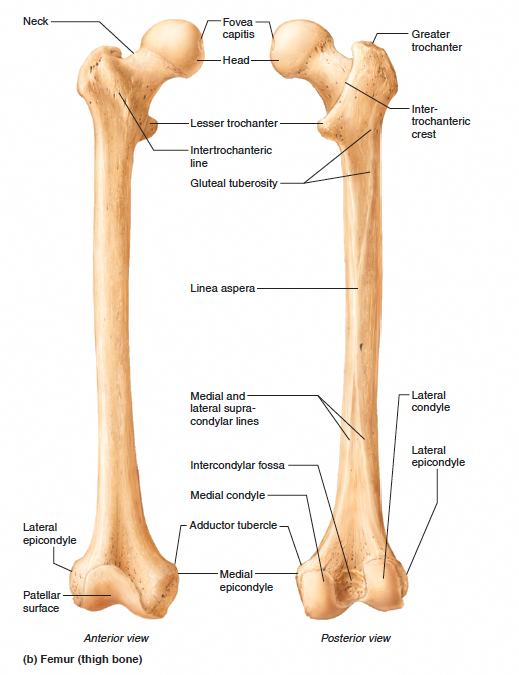
Vascular Anatomy of the Thigh Vascular Anatomy in the Femoral Region. On the lateral aspect of the thigh the fascia lata thickens to form the iliotibial tract. The single bone in the thigh is called the femurThis bone is very thick and strong due to the high proportion of bone tissue and forms a ball and socket joint at the hip and a modified hinge joint at the knee.
It is the junction of the thigh and the leg and is a hinge joint. Most of the thigh muscles are contained within two large groups of muscles referred to as the quads which extend the leg at the knee and the hamstrings which flex the leg at the knee. The superficial layer is attached to the iliac crest and descends lateral to the tensor fasciae latae muscle.
Its the area that runs from the hip to the knee in each leg. Also called the thigh bone this is the longest bone in the body. Anterior medial and posterior.
The thigh is best described in terms of compartmental anatomy and is composed of anterior posterior and medial adductor compartments. 5 Adductor longus muscle. These are the gluteus maximus gluteus medius gluteus minimus and tensor fasciae latae.
3 Rectus femoris muscle. 430 is the most superficial muscle on the medial side of the thigh. 6 Adductor brevis muscle.
It arises by a thin aponeurosis from the anterior margins of the lower half of the symphysis pubis and the upper half of the pubic arch. Medial thigh musclesAdductor longus muscleAdductor magnus muscleAdductor. It is also known as the calf bone as it sits slightly behind the tibia on the outside of the leg.
A hinge joint bends back and forth in one plane unlike the ball-and-socket joint of the hip. Legs are used for standing and all forms of. 9 Gluteus maximus muscle.
2 Vastus medialis intermedius muscles. Human Muscles July 20 2016. Large and superficial muscles which mainly abduct and extend the thigh at the hip joint.
Muscles of the Thigh - Anterior - Medial - Posterior - TeachMeAnatomy. Each compartment has a distinct innervation and function. The musculature of the thigh can be split into three sections.
The knee joint is commonly injured so understanding its anatomy can help you understand the conditions that cause problems so you stay safe and prepared. In human anatomy the thigh is the area between the hip and the kneeAnatomically it is part of the lower limb.
 Thigh Muscles Side View Human Muscle Anatomy Human Anatomy Leg Anatomy
 Leg Muscle Diagram Anatomy Of The Thigh Very Best Example Image Gallery Leg Muscles Human Muscle Anatomy Leg Muscles Anatomy Muscle Diagram
 Superficial And Deep Muscles Of The Thigh Leg Muscles Anatomy Muscle Anatomy Leg Muscles
 3d Printing Videos Vase 3dprintercraftsproducts Human Anatomy And Physiology Human Body Anatomy Anatomy Bones
 Thigh Muscle Diagram Leg Muscles Diagram Leg Muscles Anatomy Muscle Diagram
 How To Grow A Pair Thigh Muscle Anatomy Thigh Muscles Leg Muscles Anatomy
 Hip Thigh Atlas Of Anatomy Human Body Anatomy Muscle Anatomy Human Anatomy
 Hip Thigh Atlas Of Anatomy Human Muscle Anatomy Human Body Anatomy Muscular System Anatomy
 Top 8 Exercises To Build The Body Of A Greek God Leg Muscles Anatomy Muscle Anatomy Body Anatomy
 Leg Anatomy Nerve Anatomy Muscle Anatomy Medical Anatomy
 Mendmeshop Com Hamstring Injuries Muscle Anatomy Hamstring Muscles Leg Muscles Anatomy
 Hip Thigh Atlas Of Anatomy Human Body Anatomy Human Muscle Anatomy Muscle Anatomy
 Vaughan S Blog The Muscles In The Leg Muscle Anatomy Body Anatomy Human Body Anatomy
 Nerves Of The Legs Jpg 3000 2250 Inner Thigh Muscle Leg Muscles Anatomy Thigh Muscles
 Hip And Thigh Anatomy Thigh Muscle Anatomy Thigh Muscles Muscle Diagram
 Groin Muscles Diagram Koibana Info Thigh Muscle Anatomy Muscle Anatomy Hip Anatomy
 Thigh Human Anatomy Organs Anatomy Bones Physiology Anatomy


Post a Comment for "Anatomy Of The Thigh"