Important Light Bending Structure Of The Eye
The size of the pupil which controls the amount of light entering the eye is adjusted by the iris dilator and sphincter muscles. The lens bends light rays so that they form a clear image at the back of the eye - on the retina.
 Light Clipart Reflection Curve Mirrors Refraction Lenses Curved Mirror Refraction Convex Mirror
Most refraction in the eye occurs when light rays travel through the curved clear front surface of the eye cornea.

Important light bending structure of the eye. Click card to see definition. The colored ring of tissue behind the cornea that regulates the amount of light entering the eye by adjusting the size of the pupil. The transparent structure suspended behind the iris that helps to focus light on the retina.
The cornea provides about two-thirds of the power of the eye owing to the fact that speed of light changes considerably while traveling from air into cornea. The lens is a transparent body behind the iris the coloured part of the eye. Tap again to see term.
Fluid filling the anterior segment of the eye. The eyes natural lens also bends light rays. Tap card to see definition.
The iris coloured part of the eye is the structure that will contract or dilate to make the pupil larger or smaller and therby regulate the amount of light entering the eye. Associated with the eyelids are both typical sebaceous glands and modified sebaceous lands called ____ that help to lubricate the eye. Why is important to.
Composed of toughwhite connective tissue. The iris coloured part of the eye is the structure that will contract or dilate to make the pupil larger or smaller and therby regulate the amount of light entering the eye. Pupil regulates and controls the amount of light entering the eye.
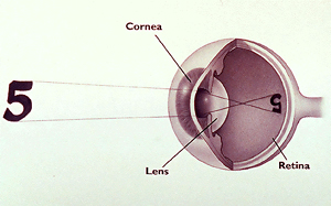
The cornea and lens both help to do this. The surface of the cornea is where light begins its journey into the eye. It primarily provides a fine-tuning adjustment to the primary focusing structure of the eye which is the cornea.
Since refraction is the measurement of how light rays bend when they enter the lens of the eye a refraction test looks at the degree to which light bends as it moves through the cornea and the lens. Transmits sound vibrations to the ossicles. Inflammation of the conjunctiva characterized by redness itching often accompanied by a discharge.
The cornea gives the initial bend to the light but the lens is the fine tuner. As the lens is. Trace the pathway of light through the eye to the retina.
State one function of pupil in human eye. Certain eye structures have refractive properties similar to water or lenses and can bend light rays into a precise point of focus essential for sharp vision. The ray diagram in Figure 2 shows image formation by the cornea and lens of the eye.
State one role of ciliary muscles in the human eye. The cornea is the transparent window in this white sac which allows the objects you are looking at to be carried in the form of light waves into the interior of the eye. The iris is the pigmented circular structure concentrically surrounding the center of the eye the pupil which appears to be black.
Click again to see term. So the images we see are reversed from left to right upside down and smaller than the object. Ciliary muscles help the eye lens to focus the image of an object on the retina by increasing or decreasing the curvature of eye lens.
The anterior aspect of each eye is pritected by the _____ which have eyelashes projected from their edges. Click card to see definition. Attaches the lens to the ciliary body.
The eye is enclosed by a tough white sac the sclera. The lens can change shape with the help of the ciliary body which contains fine muscle fibres that pull on it. Refraction or the bending of light inside the eye occurs when light travels through lens.
The lens then focuses images on the retina. Some doctors just shine a light into the patients eyes examining how much light bounces off the retina. The light bends either outward or inward depending on the lens.
Structures in the eye bend the light rays entering the eye so that when they reach the retina they are focused. If these light rays are not brought back together in precisely the same places a blurred image results. Why is important to.
Transmits the vibratory motion of the stirrup to the fluid in the inner ear hammer malleus auditory tube. The image formed on the retina is a result of the light bending activity of the lens. Tap card to see definition.
This bending of the light is important since all light originating from a given point source in the outer world must converge on a single spot on the photoreceptive surface of the eye the retina in order for a focused image to be perceived. Three circular passages each in a different plane of space. Contains muscle that controls the size of the pupil.
The rays bend according to the refractive indices provided in Table 1.
 This Item Is Unavailable Etsy Hanging Lamp Lamp Make A Lampshade
 The Structure Of The Eye And The Functions Of These Accessory Structures Eye Structure Ciliary Muscle Eyes
 Eye The Human Eye And The Focus Vision Structure And Functioning Animate Structure And Function Human Eye Animation
 Vision Boundless Biology
 Refractive Index Definition Equation Physics Physics Formulas School Study Tips
 Tesseract 7 Glow Light Installation Light Art Installation Light Sculpture
 Answers To This Module
 Ten Truss Bridge Google Search Roof Truss Design Roof Trusses Roof Structure
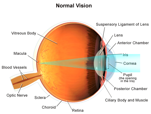 Normal Vision
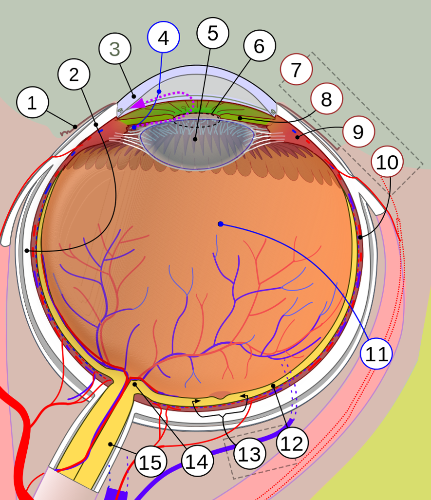
 Unit 5 Eye Flashcards Quizlet
 Eye Anatomy And How The Eye Works Davidson Eye Associates
Ledbysource On Instagram The Pineal Gland Is Nestled In The Center Of Our Brains Between The Two Hemispheres With A Sim Pineal Gland Gland Mind Body Spirit
 Crista Ampullaris Start Of Semicircular Canals Detect Angular Acceleration At Each Ampullae There Is Hair Cells With Cilium And S Cabeca E Pescoco Pescocinho
 Ametropia Hyperopia Myopia Astigmatism Presbyopia Rayur Astigmatism Eye Exam Presbyopia
 Pin On Refraction And Lenses
 Lenses Are Important In Scattering Converge Diverging Light As Needed To Correct Vision In Hyperopia A Convex Lends Bends The Chapter 16 Focus Light To Focus
 The Origin Of Courti Within The Cochlear Duct Contains Epithelia Cells With Stereocilia And Are Connected To Neuronal Str Holistic Medicine Sensory Membrane
 Photoreceptor Cells Consist Of Rod And Cone Cells Rod Cells See Dark Light Rod Cells Have Rhodopsin Opsin Protein Portion R Neurons Color Light In The Dark
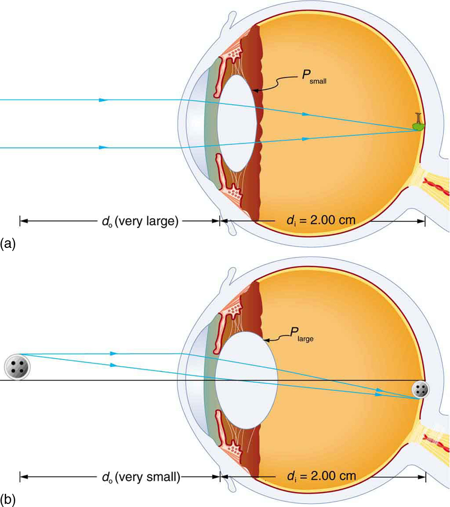 Physics Of The Eye Physics
Post a Comment for "Important Light Bending Structure Of The Eye"